|
 CF-1
CF-1
Digital Retinal Camera |
PDF |
|
Get the best images for the best
diagnosis—quickly, easily and
comfortably In terms of
image quality and workflow
efficiency, Canon’s digital
retinal cameras are in a class
of their own. Every aspect of
image capture—from
the precision retinal imaging
optics to the advanced digital
SLR technology—has been
developed in-house to create a
seamless, total imaging system
that provides unsurpassed
diagnostic image quality and
speed. The CF-1 takes this a
step further with an all-new
ergonomic design that makes it
easier and more comfortable than
ever to achieve precise
images during color/red-free
imaging and fluorescein
angiography. |
 |
|
|
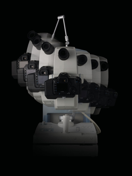
Industry-leading image quality
The CF-1 integrates a digital
SLR camera from Canon’s renowned
EOS series equipped with a
high-sensitivity CMOS image
sensor that achieves ultra-high
resolution images with superior
detail, contrast, and color
fidelity. The sensor’s high pix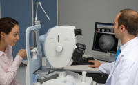 el
count ensures that images are
sharp and clear even when
enlarged, and its high
sensitivity reduces the amount
of light needed for image
capture, a factor that helps
increase patient comfort during
examinations. After capture,
images are transferred to the
connected PC for immediate el
count ensures that images are
sharp and clear even when
enlarged, and its high
sensitivity reduces the amount
of light needed for image
capture, a factor that helps
increase patient comfort during
examinations. After capture,
images are transferred to the
connected PC for immediate
on-screen observation. |
|
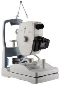
Intuitive, comfortable
operation The
streamlined, ergonomic design of
the CF-1 is not only inviting in
appearance, it makes the camera
a pleasure to operate. It’s
easily adjustable to comfortably
accommodate each examinee.
Smooth, precise pan and tilt
movement allows you to easily
achieve desired views of the
retina without requiring the
examinee to move their gaze.
Alignment and focus is easy as
well, helping you to complete
exams in less time.
Easy positioning with smooth pan
and tilt movement |
 |
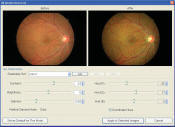
2x digital magnification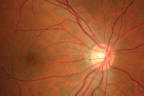
With one push of a button on the
CF-1 control panel, you can get
a closer, more enlarged view of
the retina for immediate review
on the connected PC. “2x mode”
works by automatically cropping
out the peripheral edges of the
image so that the region of
interest is larger in the frame,
making it quick and easy to
confirm image focus and other
factors. The end result looks
the same as optical
magnification yet is achieved in
a fraction of the time.
Close-ups are exceptionally
clear and detailed thanks to the
high pixel count of the
integrated digital SLR camera. |
|
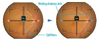
Easy alignment and focus
Get clear, sharply-focused
images every time in two simple
steps. First, align the two
halves of a split line using the
focusing knob to bring the image
into focus. Then adjust the
working distance to avoid flash
flare by shifting the joystick
until the two side dots are
clear in the viewfinder. |
 |
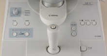
Ergonomic control panel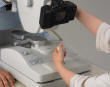
Controls for key features—such
as shutter release, lamp
setting, ISO adjustment, and
mode switching—are grouped
together for easy one-handed
operation in darkened rooms.
|
 |
|
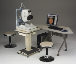
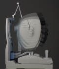
Compact, streamlined design
All CF-1 functions are
integrated into a single,
streamlined tabletop unit with a
power supply that’s built into
the base. The new co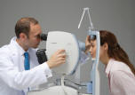 mpact
design enables closer
face-to-face interaction with
the examinee and allows easy
access to the examinee’s eye.
And the detachable digital
camera conveniently receives its
power through the body of the
CF-1, so there’s no separate AC
adapter to plug in and cables
are kept neatly tucked out of
the way. mpact
design enables closer
face-to-face interaction with
the examinee and allows easy
access to the examinee’s eye.
And the detachable digital
camera conveniently receives its
power through the body of the
CF-1, so there’s no separate AC
adapter to plug in and cables
are kept neatly tucked out of
the way.
Allows easy access to the
examinee’s eye |
|
Motorized chin rest
The motorized chin rest can be
moved up and down to accommodate
the examinee's height using a
pair of buttons
located on the unit's control
panel. |
|
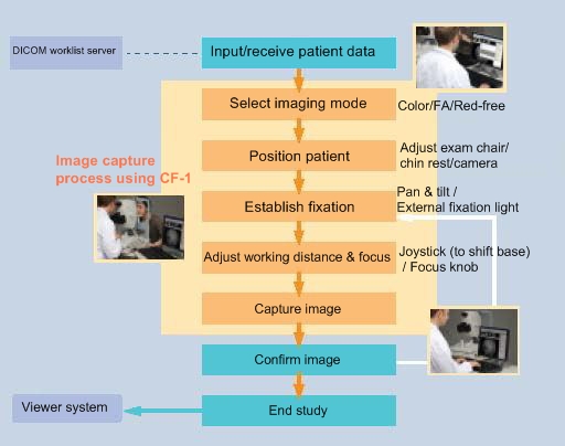
Streamlined system and workflow
The efficiency of the CF-1 goes
beyond image capture. The
network capability and control
software of the CF-1 work to
streamline the entire diagnostic
workflow, allowing you to
conveniently review, analyze,
print, store and even transmit
images to remote viewing
locations. The DICOM-compliant
network interface enables easy
integration with existing image
management systems and
allows connection to a
variety of network
configurations such as LAN or
WAN and PACS communication. |
|
Simple, straightforward control
software The
bundled Retinal Imaging Control
Software for the CF-1 puts tools
for comprehensive study
management and image capture
control at your fingertips,
in an intuitive graphical
interface that’s simple and
straightforward to use. The
PC-based software provides
quick, easy input and access to
information and images required.
A typical exam begins with the
input of patient data, which can
be automatically received from a
DICOM worklist server or
manually entered. When the
shutter-release button of the
CF-1 is pressed, images are
captured using the ideally
 set
parameters of the attached
digital SLR camer set
parameters of the attached
digital SLR camer a
and displayed on the PC monitor
for immediate review. The
software’s preview tools include
image magnification and
adjustment controls for
contrast, brightness, and other
factors to aid in image
confirmation. When the study is
finished, saved images can be
printed and a
and displayed on the PC monitor
for immediate review. The
software’s preview tools include
image magnification and
adjustment controls for
contrast, brightness, and other
factors to aid in image
confirmation. When the study is
finished, saved images can be
printed and
automatically transferred to a
viewer system. |
| |
Workflow of a typical exam |
|
|
Features
The CF-1
integrates a digital SLR camera from
Canon’s EOS series to achieve high
resolution images with excellent detail,
contrast, and color fidelity.
•
Intuitive, comfortable operation
Precise pan and tilt movements allow
the operator to easily achieve views
of the retina while reducing the
need for patients to change their
gaze.
•
50-degree angle of view
The 50-degree view angle enables
easy alignment and focus.
• 2x
digital magnification
Zoom the retina to 2x magnification
and the system automatically crops
out the peripheral edges of the
images.
•
Ergonomic control panel
Operator controls are grouped
together for one-handed operations
in darkened exam rooms.
|
Specifications
|
General |
|
|
|
Types of
Imaging |
Color,
red-free and Fluorescein
angiography imaging. Stereo
assist device (optional) |
|
|
Angle of
view |
50
degrees, 43 degrees (2x digital
magnification) |
|
|
Minimum
pupil size |
5.2 mm
(in SP mode 4.3 mm) |
|
|
Patient’s
diopter |
Without compensation lens, -10D
to +15D
With “-“ compensation lens, -7D
to -31D
With “+” compensation lens, +11D
to +33D |
|
|
Light
Source |
Halogen lamp for observation,
xenon tube for photography |
|
|
Eye
fixation lamp |
External type standard, internal
type optional |
|
|
|
Adjustments |
|
|
|
Base
Adjustments |
Forward/backward 65 mm
Right/left 110 mm
Up/down 30 mm |
|
|
Working
Distance |
35 mm |
|
|
Focus
adjustments |
Split
lines focusing, working distance
dots adjustment |
|
|
Panning
Range |
Right/left 30 degrees |
|
|
Tilting
Range |
Upward
15 degrees
Downward 10 degrees |
|
|
|
Electrical and Environmental |
|
|
|
Power
Supply |
AC
100-240V 50/60 Hz 7-3A |
|
|
Power
Consumption |
Normal
approx. 100 VA
Manixmum 720 VA |
|
|
Operating
Environment |
Temperature 10 degrees C to 35
degrees C
Humidity 30” RH to 80% HR
(non-condensing) |
|
|
|
Physical Characteristics |
|
|
|
Dimensions
(W x D x H) |
12.6
in. x 10.9 in. x 22.3 in.
320 mm x 531 mm x 566 mm |
|
|
Weight |
Approx. 57 lbs. (26 kg) |
|
|
|